What Is a Skin Analyzer and How Does It Work?

Time:2026-11-03 Author:Henry
0%

A mirror can show a dry patch or a shiny forehead, but it cannot reveal every detail of skin health. A Skin Analyzer uses cameras, lighting, and software to assess visible features such as pores, redness, texture, and uneven pigmentation. Some devices capture several images under different types of light. Others use contact sensors to estimate moisture or oil levels. The exact measurements depend on the model and its testing method. Small details matter. Even room lighting, makeup, and recent skincare can affect a scan.

The results can help people notice patterns over time and give skincare professionals a clearer starting point for discussion. A screen might highlight areas that are difficult to see in ordinary light, then display scores or color maps. Those numbers are estimates, not a complete medical assessment. No scan tells the whole story. Skin changes with age, weather, hormones, and daily habits, so a single reading can be misleading. It is worth asking how a device collects data and what its results actually mean. For persistent irritation, changing spots, or other concerns, a qualified healthcare professional can provide an appropriate evaluation. Used thoughtfully, a Skin Analyzer can support informed conversations, while leaving room for human judgment and a little healthy doubt.
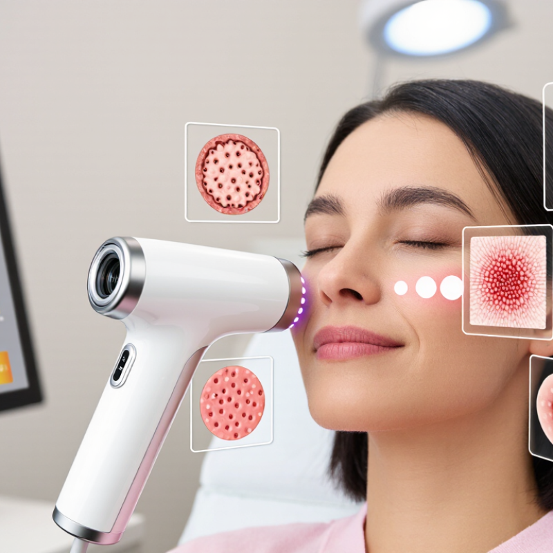
What Is a Skin Analyzer and How Does It Work?

Definition and Purpose of a Skin Analyzer

A skin analyzer is a digital assessment tool that helps examine visible and less obvious features of the skin. It can record pores, oiliness, redness, pigmentation, texture, and fine lines under consistent lighting. Depending on its design, it may use close-up photography, cross-polarized light, or ultraviolet imaging. The images are processed into maps or scores, giving a practitioner clearer areas to inspect. It is an aid, not a diagnosis. That distinction matters.

Its main purpose is to establish a baseline and make consultations more specific. A clinician might compare cheek redness or forehead oiliness across visits, rather than relying only on memory. The images can also help explain why a routine is adjusted, or identify changes worth discussing. But scores are not universal, and they should not be treated as a measure of overall health.

Lighting, recent exercise, makeup, dehydration, and even camera position can affect the reading. A dry room may make skin appear different from a humid one. Results should therefore be interpreted alongside questions about symptoms, habits, and medical history. One scan cannot tell the whole story. Thoughtful use means noting uncertainty, repeating images under similar conditions, and referring persistent or concerning changes to a qualified health professional.

Key Sensors and Imaging Components

A skin analyzer combines controlled lighting, image sensors, and measurement software. Its camera usually captures visible-light images, while cross-polarized illumination can reduce surface glare. Some systems add ultraviolet or multispectral light to reveal features that ordinary photographs may miss. The sensor records reflected light; software then compares color, texture, pores, or visible spots across regions. Small details matter: a warm lamp can shift redness, and a tilted chin can alter shadows. Not magic.

Sensor quality is only part of the measurement. Calibration, camera distance, exposure, and repeatable positioning all affect results. The International Commission on Illumination’s CIE 15:2018 guidance explains how controlled viewing conditions support consistent color measurement. For context, a 2018 Cochrane review reported dermoscopy sensitivity of about 92% at a specified 80% specificity threshold for melanoma assessment; that clinical result does not validate cosmetic analyzer scores. It does show why image interpretation needs context, not just a sharp picture.

A practical setup uses diffuse white LEDs, a fixed chin rest, and a color reference target. Some devices capture polarized and non-polarized images separately, helping distinguish surface reflection from deeper color patterns. Yet algorithms can still misread freckles, facial hair, or uneven lighting. Results are estimates, not diagnoses. I would question any score that hides its lighting conditions or calibration method.

How Skin Data Is Captured

A skin analyzer captures information through a camera, sensor, or both. Before a scan, the device may ask you to remove makeup and sit in a fixed position. You place your face near the camera, where steady lighting helps reveal visible details. Some systems take several images, including close-ups of the cheeks, forehead, and nose. Others measure features such as surface moisture or oil levels using contact sensors. Small changes in distance or lighting can affect the reading.

Software then examines the captured data for patterns, such as visible pores, redness, uneven tone, or fine lines. The result is an estimate, not a diagnosis. A bright room, recent exercise, or a newly applied moisturizer may change what the device records. Even a clear score can miss context, such as sensitivity that appears only after a certain product. It is worth asking how the device captures data and whether results can be compared consistently over time.

Tips: Scan clean, dry skin under similar lighting each time. Keep your face at the same distance, and avoid comparing readings taken after exercise with morning readings. Note recent skincare changes beside each result. The numbers may look precise, but they still need thoughtful interpretation.

How the Analyzer Interprets Skin Features

A skin analyzer uses a camera, lighting, and sometimes sensors to record visible skin features. The person sits still while the device captures close-up images, often under consistent illumination. Software then identifies patterns such as pores, surface shine, redness, uneven tone, and fine lines. Some systems estimate moisture or oil levels, but their methods vary.

The analyzer interprets these features by comparing image details with programmed reference ranges. For example, it may count visible spots or measure how evenly light reflects from the skin. The results are estimates, not a medical diagnosis. A score can change with room lighting, makeup, recent exercise, or even camera position. That matters. One scan cannot explain every change in someone’s skin, and the software may miss context a trained professional would notice.

Tips: Cleanse gently before a scan and avoid applying makeup or rich creams. Keep your face relaxed and follow the same positioning each time. Compare results under similar conditions, rather than treating one number as a final verdict. Skin is variable. A little uncertainty is normal.

What Is a Skin Analyzer and How Does It Work? - How the Analyzer Interprets Skin Features
Skin Feature How It Is Captured or Measured How the Analyzer Interprets It Important Limitations
Surface texture and visible pores Standardized photographs use visible light to record skin-surface detail. Image-processing software may assess local contrast, texture patterns, and the apparent size of pore openings. The system identifies visible texture patterns and estimates where pores appear more prominent in the captured image. Lighting, camera distance, focus, makeup, and image resolution can change the result. A photograph does not directly measure pore depth or diagnose a skin condition.
Fine lines and wrinkles Images are analyzed for narrow lines, shadows, and changes in surface contrast; some systems compare multiple facial regions or images over time. Detected line patterns may be grouped by location, visibility, or estimated prominence to support a cosmetic skin assessment. Facial expression, hydration, lighting direction, and image angle can affect how lines appear. Results are estimates, not a clinical measurement of skin aging.
Visible redness and uneven tone Color photographs are evaluated for differences in color and brightness across areas of the face. Some devices use controlled illumination to improve consistency. The analyzer maps visible color variation and may flag areas that look redder or less even than surrounding skin. Skin tone, white balance, ambient light, and recent exercise or temperature changes can affect color readings. Redness analysis cannot determine its medical cause.
Pigmented spots and dark marks Image analysis identifies localized areas that differ in color or contrast from nearby skin. Some systems use additional illumination modes to make certain surface patterns easier to see. Spots may be counted, mapped, or compared by visible appearance to help track cosmetic changes over time. Image-based analysis cannot determine whether a spot is harmless or medically concerning. New, changing, or unusual spots should be assessed by a qualified healthcare professional.
Oiliness and surface shine Some analyzers assess reflected light in facial images; dedicated instruments may estimate surface sebum using a measurement strip or sensor. Reflection patterns or sebum readings can indicate areas that appear oilier, often around the forehead, nose, and chin. Shine can also result from lighting, skincare products, sweat, or recent cleansing. A visual estimate is not equivalent to a direct sebum measurement.
Skin hydration Dedicated probes may estimate water content in the outer skin layer by measuring electrical properties such as capacitance. A standard camera image cannot directly measure hydration. The instrument converts its sensor reading into an estimate that can be compared across sites or repeated sessions under similar conditions. Readings vary with location, room conditions, recent washing, and product use. Hydration scores are device-dependent and should not be treated as a diagnosis.
UV-related surface patterns Some systems use ultraviolet or other specialized illumination to capture patterns not readily visible in ordinary photographs. The software highlights contrast patterns that may be associated with accumulated sun exposure or pigmentation near the skin surface. These images are screening or cosmetic visualization tools, not a direct measure of total UV damage or a substitute for a medical skin examination.
Overall skin scores and progress tracking Software combines selected image features or sensor readings, sometimes comparing results with reference ranges or earlier measurements. A summary score can make repeated observations easier to review and may help track visible changes when images and conditions are consistent. Scoring methods differ between devices. Scores depend on capture quality and the system’s algorithms, and should not be interpreted as a medical diagnosis or guaranteed prediction.

Common Uses and Limitations of Skin Analysis

A skin analyzer uses a camera and sensors to estimate visible features such as pores, surface oil, redness, and uneven pigmentation. Some devices also assess moisture or texture. The results can help a skincare professional discuss concerns with a client, such as dry patches around the cheeks or shine across the forehead. Useful, but not definitive.

Commonly, these tools support consultations and track changes over time. A professional may compare images taken under similar conditions, then adjust a routine or suggest when medical advice is appropriate. For a fair comparison, the face should be clean, makeup-free, and photographed with consistent lighting. Even then, readings can vary.

Skin analysis has real limits. Lighting, camera quality, recent product use, and hydration can affect results. Skin tone may also influence how some imaging systems detect redness or discoloration. An analyzer cannot confirm a skin disease or replace an examination by a qualified clinician. Its scores may look precise, but precision is not the same as certainty. I would treat a reading as a conversation starter, not a verdict.

What Is a Skin Analyzer and How Does It Work?

Skin analyzers use images and other measurements to assess visible features such as pores, pigmentation, redness, and wrinkles. Some systems use different light wavelengths to reveal different skin patterns.

The chart shows conventional wavelength bands: visible light is approximately 400–700 nm, UVA is 315–400 nm, and UVB is 280–315 nm. These are spectral definitions, not specifications for every analyzer. Image-based results are estimates and cannot diagnose skin conditions; lighting, device settings, and skin characteristics can affect them.

FAQS

What does a skin analyzer measure?

It records features such as pores, oiliness, redness, pigmentation, texture, and fine lines. Some devices also estimate surface moisture or oil levels.

Can a skin analyzer diagnose a condition?

No. It is an assessment aid, not a diagnosis. Persistent or concerning changes should be discussed with a qualified health professional.

How does a skin analyzer capture skin data?

A camera records images under controlled lighting. Some systems use polarized, ultraviolet, or multispectral light. Others use contact sensors.

What can affect scan results?

Makeup, recent exercise, dehydration, room humidity, lighting, and camera position can affect readings. Even a tilted chin can change shadows.

How should I prepare for a scan?

Use clean, dry skin and remove makeup if requested. Keep your face at the same distance each time. Small details matter.

Can I compare results from different visits?

Yes, cautiously, if lighting and positioning are similar. Note skincare changes and scan timing; a morning reading may differ after exercise.

What do analyzer scores actually mean?

Scores are estimates from images or sensor readings. They are not universal measures of skin health, and their meaning may be unclear.

What should I ask before trusting a result?

Ask about lighting, calibration, camera distance, and how images are compared. I would question any precise score that hides these details.

Conclusion

A Skin Analyzer is a tool designed to assess visible skin characteristics and turn them into organized information that can support skincare discussions. Depending on its design, it may use cameras, magnification, controlled lighting, and sensors that detect features such as surface texture, oiliness, moisture, pores, redness, and areas of uneven pigmentation. These components help capture images or readings under consistent conditions, making it easier to observe details that may be difficult to notice with the naked eye.

During an analysis, the user’s skin is positioned for imaging or measurement, and the device records relevant data. Its software then processes the images and readings, identifies patterns, and presents estimates or visual maps of selected features. Results can help track changes over time or guide general skincare choices, but they are not a medical diagnosis. Lighting, positioning, skin preparation, and device capabilities can affect accuracy, so findings should be treated as informative rather than definitive.


Henry

Henry

Henry is a dedicated marketing professional with a profound expertise in the company's offerings. With years of experience in the industry, he possesses an impressive understanding of the market dynamics and consumer behaviors that drive success. Henry is committed to sharing his insights through......