A Facial Skin Analyzer is a camera-based system that captures details a mirror may miss. Depending on the device, it uses standard, cross-polarized, or ultraviolet lighting to examine visible features such as pores, redness, fine lines, and pigmentation. Software then processes the images and presents estimates, often alongside a skin score or care suggestions. It is an assessment tool, not a diagnosis.
The interest is understandable. The American Academy of Dermatology estimates that acne affects up to 50 million people in the United States each year, while Grand View Research valued the global skincare products market at about $115.65 billion in 2024. These figures describe a large need and market, not proof that any analyzer improves skin. That distinction matters.
In practice, a user sits before a fixed camera while the device takes images under controlled lighting. An algorithm compares visual patterns and may track changes across later scans. Results can vary with lighting, makeup, camera position, and the system’s training data. A score may look precise, yet it remains an estimate. Not a diagnosis. For consumers and professionals, the useful question is not whether the device can reveal everything, but what it measures, how consistently it measures it, and where its limits lie. Those limits are easy to overlook. A clinician should assess persistent or concerning skin changes rather than relying on an image report alone.
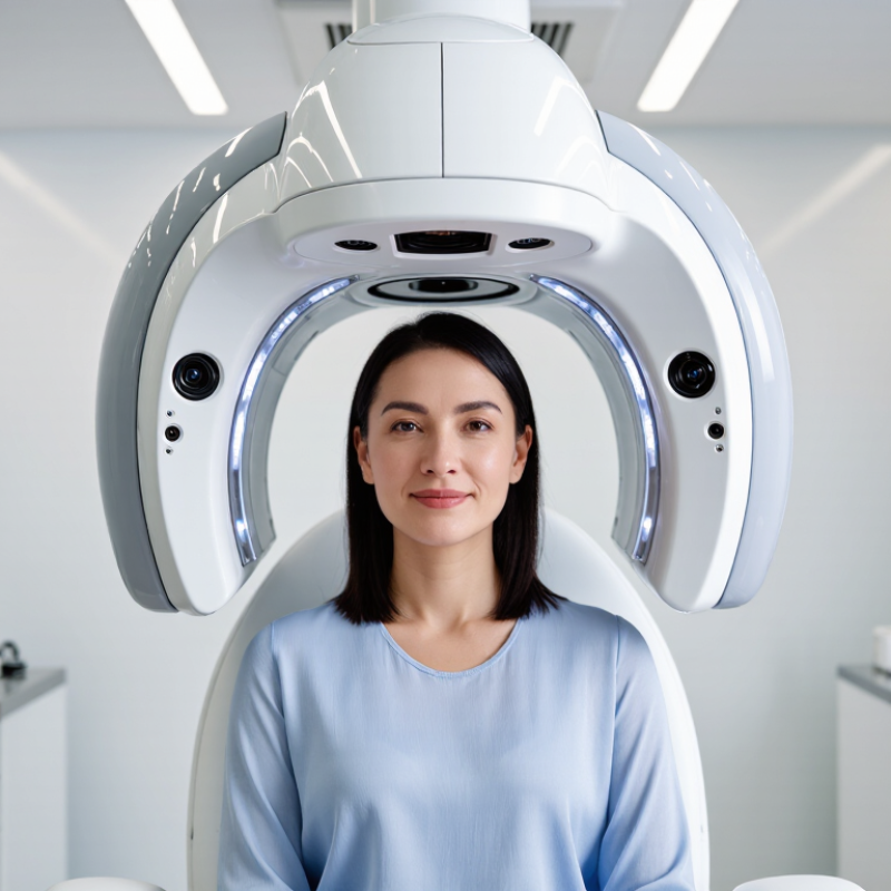
A facial skin analyzer is a device or imaging system that captures close-up pictures of the face and estimates visible skin features. Depending on its design, it may use standard or cross-polarized light to show details such as pores, fine lines, redness, oiliness, and uneven pigmentation. Some systems compare images taken under different lighting conditions. The result is a structured snapshot, not a complete account of skin health.
During a session, the user usually removes makeup and sits still while a camera records the face. Software then processes the images and displays measurements or highlighted areas. These estimates can help track changes over time, especially when lighting, distance, and facial position stay consistent. Results may vary with lighting, recent skincare, or camera settings. The numbers can look more certain than they are. An analyzer cannot diagnose a skin condition or replace advice from a qualified clinician.
Tips: Take comparison photos under similar conditions, and note recent product use. Treat small score changes cautiously. A clear image is useful, but it is not the whole story.
A facial skin analyzer uses images—often captured under different lighting—to help assess visible features such as pores, spots, redness, and wrinkles. Some devices also use probes to measure properties such as hydration or surface pH. The chart compares a representative midpoint of typical facial skin surface pH with neutral pH; actual skin pH varies.
How to read it: Facial skin surface pH is commonly reported around 4.5–5.5, so 5.0 is shown as the range midpoint. Neutral pH is 7.0. A skin analyzer’s methods and available measurements depend on the device.
A facial skin analysis system combines several parts to capture and interpret visible skin features. A high-resolution camera records areas such as the forehead, cheeks, and chin. Controlled lighting helps reveal texture, pores, redness, and uneven tone. Some systems use different light modes, including polarized or ultraviolet illumination, to highlight features that ordinary room lighting can hide.
The camera is only one piece. Light sources need consistent brightness, while positioning guides help keep the face at a repeatable distance and angle. Sensors may monitor image conditions, and software processes the photographs into measurements or visual maps. A screen then displays the results for review. Small changes matter: a stray hair, a tilted chin, or makeup can affect an image. The system does not read skin perfectly. It can support a closer conversation, but it cannot replace a qualified professional’s assessment. Calibration and clean equipment are easy to overlook, yet they influence consistency. That part is less glamorous. Even careful users may interpret a color map too confidently, so findings should be considered alongside the person’s history and an in-person examination.
| System Component | What It Does | How It Works | Typical Information Provided |
|---|---|---|---|
| Lighting and illumination | Provides controlled light so the camera can capture comparable images. | Visible-light systems commonly use diffuse, even illumination. Some systems also use cross-polarized light to reduce surface glare or ultraviolet illumination to reveal fluorescence patterns. | Visible-light views can show tone and texture; cross-polarized views can make redness and uneven pigmentation easier to assess; UV images may show certain surface fluorescence patterns. |
| Camera and image sensor | Captures photographs of the skin for visual or software-assisted assessment. | A digital sensor records reflected light as image data. Image quality depends on factors such as focus, exposure, resolution, and consistent lighting. | Photographic records of visible features such as pores, fine lines, spots, redness, and texture. |
| Optics and filters | Direct and control the light reaching the sensor. | Lenses focus the image. Optical filters may select or block parts of the light spectrum, supporting different image modes, such as polarized or UV-assisted capture. | Images that emphasize different visible or light-related features; results depend on the device’s optics and capture settings. |
| Positioning and capture station | Helps keep the face in a consistent position during image capture. | A chin rest, forehead support, distance guide, or marked positioning area can reduce changes in angle and distance between sessions. | More consistent images for comparison over time; positioning does not itself measure skin condition. |
| Image-processing software | Organizes images and may identify or quantify selected visual patterns. | Software can compare images, apply image-processing methods, and generate scores or overlays. The specific features and methods vary by system. | Possible outputs include counts or visual maps of selected features, comparison views, and summary scores. These are estimates based on captured images. |
| Calibration and quality checks | Support reliable image capture and repeatability. | Depending on the system, checks may include camera focus, exposure, lighting consistency, and use of a reference target. Following the device’s operating instructions is important. | Helps identify capture conditions that could affect image comparisons; it does not guarantee a clinical diagnosis. |
| Display and report interface | Presents images and analysis results to the user or practitioner. | A screen or report may show original images, processed views, and comparisons between visits. Interpretation should account for lighting, skin preparation, and other capture conditions. | A visual record that can support discussion of skin appearance and changes over time. |
How the system works: The device captures images under selected lighting conditions, then its software displays or analyzes those images. A facial skin analyzer provides image-based information and should not be treated as a stand-alone medical diagnosis.
A facial skin analyzer begins with a controlled image, not a diagnosis. The user rests their chin on a support while a camera records the face under set lighting. Some systems capture ordinary light; others use polarized or ultraviolet light to reveal different surface patterns. Small details matter. Stray hair, makeup, and a tilted head can affect what the camera sees.
The software divides the image into facial areas, such as the forehead, cheeks, and nose. It then measures visible features, including pores, fine lines, redness, and uneven pigmentation. Depending on the device, sensors may also estimate surface oil or moisture. These readings are compared with internal reference ranges and converted into images, scores, or trend charts. A bright spot on a screen is a measurement cue, not proof of a medical condition.
Consistent capture makes comparisons more useful. Keeping the same distance, lighting, and facial expression can help show changes over time. Still, skin tone, room conditions, and image quality may influence results. Not magic. A polished report can look more certain than the underlying data really is. The analyzer offers a structured snapshot; a trained professional can add context when a finding is unclear or persistent.
A facial skin analyzer uses a camera, sensors, or both to record visible skin features and estimate less obvious ones. Common readings include hydration, oiliness, pore appearance, texture, fine lines, redness, and uneven pigmentation. Some devices also use specialized lighting to reveal spots beneath the surface. The exact features depend on the device, so two systems may not report identical results.
Hydration readings can suggest whether skin looks dry, while sebum measurements estimate surface oil. Pore and texture images may show enlarged-looking openings, rough patches, or small lines around the eyes. Pigmentation tools assess the distribution of darker areas, and redness maps can highlight visible color differences. Lighting matters. Makeup, recent exercise, room temperature, and even a damp face can affect a scan. For a more useful comparison, measurements should be taken under similar conditions and interpreted alongside a person’s concerns and skin history.
A single score is only a snapshot, not a diagnosis; neat charts can hide uncertainty, and that limitation deserves attention.
A facial skin analyzer converts images into estimates of features such as visible spots, pores, texture, and fine lines. Its numbers depend on the camera, lighting, software, and image conditions. Read a score as an observation, not a diagnosis. Compare scans only when conditions are similar. Keep the same room, distance, and time of day; avoid comparing a bright bathroom selfie with a dim clinic scan. Small shifts in light can make pigmentation look stronger or weaker.
That matters. The International Skin Imaging Collaboration’s 2020 challenge dataset contained 33,126 dermoscopic images. Those close-up lesion images are not the same as consumer facial scans, so their results cannot validate every analyzer. A device’s percentage may look exact, but it can reflect its own image library and scoring rules.
Not a diagnosis. Treat persistent redness, pain, or changing marks as reasons to seek a qualified clinician’s opinion. I may still be tempted to trust a crisp number too much; a repeat scan can help show a trend, but it cannot explain what caused it.
It combines a camera, controlled lighting, positioning guides, sensors, software, and a display. Each part can affect the final image.
The person rests their chin on a support while the camera photographs their face. Set lighting helps reveal surface details.
Depending on the device, it may assess pores, texture, fine lines, redness, pigmentation, surface oil, and hydration.
No. Its images and scores are measurements, not a diagnosis. A qualified professional can interpret unclear or persistent findings.
Different light modes can reveal different patterns. A tilted chin may change the camera’s view.
Makeup, stray hair, recent exercise, room temperature, and damp skin may influence readings. Small details count.
Keep lighting, distance, facial expression, and room conditions similar for each scan. One score is only a snapshot.
Not entirely. Polished charts may appear more certain than the underlying data. That deserves a second look.
A Facial Skin Analyzer is a device that uses imaging and sensors to assess visible features of the skin. A typical system includes a camera or optical sensor, controlled lighting, image-capture software, and an analysis program. Together, these components help create consistent images and identify patterns that may be difficult to notice through casual observation.
During an analysis, the device captures skin images under selected lighting conditions and processes them to estimate features such as pores, fine lines, uneven tone, redness, oiliness, and moisture-related indicators. Results are usually presented as measurements or visual maps that can help users understand their skin and track changes over time. However, readings are estimates, not medical diagnoses. Lighting, image quality, skin preparation, and individual differences can affect the results, so findings should be interpreted cautiously and considered alongside professional advice when needed.

Aidiesin